“Bunker busting” Antibody-antibiotic-conjugates (AACs) successfully used to target MRSA bacteria hiding inside the host’s cells

One of the difficulties in combating MRSA is that (Staphylococcus aureus) bacteria have the ability to live inside the host’s cells where they are effectively sheltered from the action of systemic antibiotics – it is this reservoir of infection that provides the seed for the relapses that are characteristic of MRSA.

Staphylococcus aureau
Magnification 20,000

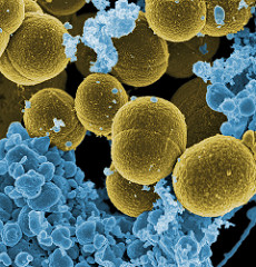
S. aureus bacteria escaping a white blood cell, x20,000 mag.
Credit: NIAID – Creative Commons CCBY2

A large team of researchers from Genentech in the USA and Symphogen in Denmark has developed a method to destroy these intracellular S.aureus bacteria that would otherwise be protected from antibiotics. The team used a novel conjugate of the antibiotic rifalogue together with monoclonal antibodies. These antibody-antibiotic-conjugates (AACs) are specifically immuno-targeted to S.aureus. The AACs remain inactive in the bloodstream, and only become active in the presence of the bacteria inside the host´s cells.

Once the AACs are taken into the host’s cells they are transported deeper – into the phagolysosomes which enclose the bacteria inside the cell. The phagolysosomes contain a proteolytic environment, and the protease enzymes found there cleave a small peptide group from the AACs – activating them. The active AACs are then able to bind to the surface of the bacteria effectively delivering their antibiotic payload to the infection’s hidden “bunker”.

Infected mice were treated with the AACs, and the team found this new treatment was much more effective than a systemic antibiotic (vancomycin). This work confirms the importance of intracellular S.aureus as a reservoir of MRSA infection, and raises the exciting possibility that these AACs might eventually be used to treat humans. The targeted approach of AACs would avoid damaging the patient’s beneficial microflora, and if their use becomes routine it would probably reduce the rate at which bacteria in general evolve resistance to any one particular antibiotic.

(This study was published in Nature:
Sophie M. Lehar et al. Novel antibody–antibiotic conjugate eliminates intracellular S. aureus, Nature (2015). DOI: 10.1038/nature16057 http://www.nature.com/nature/journal/v527/n7578/full/nature16057.html )

Child’s life saved from leukemia in ground-breaking use of gene-edited immune system cells

Doctors at Great Ormond Street Hospital (GOSH) successfully used “off the shelf” genetically engineered white blood cells (T-cells) in a last ditch effort to treat a one-year old girl, called Layla, who was suffering from acute lymphoblastic leukemia (ALL) that had resisted chemotherapy. This is the world’s first instance of this targeted cancer therapy in a human patient.

To achieve this GOSH doctors worked with research scientists at University College London’s (UCL) Institute of Child Health (ICH) and biotech company Cellectis. The gene-edited T-cells were modified using a “molecular toolkit” that scientists have pirated from a few genes found in certain bacteria – especially a biological editing tool called TALEN.
TALEN is a combination of a modular protein (TAL) that can effectively be “programmed” to find and bind very specific DNA sequences, together with an endonuclease (EN) which is a protein that can cut DNA, ready to replace that gene with the version desired.

The modified T-cells are called UCART19 cells, and they are produced to fight leukemia in a two step process:
First, they have a gene that programs for a characteristic cell surface protein deleted – so the UCART19 cells will be “invisible” and remain safe from the anitibodies that are given to leukemia patients to destroy their existing, diseased immune system.
Secondly, the T-cells have the gene for the CAR19 surface protein added – CAR19 will bind the UCART19 cells to a different protein called CD19, which is only found on the surface of immature white cells (called “blasts” – lymphoblasts in ALL) that proliferate in leukemia and “crowd out” other healthy blood cells, thus causing the disease symptoms. Once bound to the leukemia cells (lymphoblasts) the UCART19 cells recognise them as foreign and destroy them.

nci-vol-4345-72LeukemiaPubDomCredNCIAlanHooofringColourInvert
(Above: The blood stream of a healthy subject vs. a leukemia patient.
RBCs = Red Blood Cells. WBCs = White Blood Cells.

Public domain image, credit: NCI, Alan Hoofring.
Modified by J.Overton)

Clinical trials taking place at the moment normally begin with white blood cells taken from the patient because these run least risk of causing auto-immune problems, but this “bespoke” method of production is expensive. However, due to the chemotherapy and highly agressive nature of the leukemia she suffered, little Layla did not have enough white blood cells left to work with, so the team gave her “off the shelf” UCART19 cells created from donated T-cells.

Previously, this experimental treatment had only been tested on mice in the lab, in fact it was so new that GOSH had to convene an emergency ethics meeting to decide whether Layla should receive it. As routine chemotherapy and a bone marrow transplant had already failed to help Layla, and her condition was worsening, all the doctors had left to offer was either palliative care to relieve her suffering during terminal illness, or the hope of possible recovery with the UCART19 cells. So, together with Layla’s parents, they decided to opt for treatment.

After about two weeks of receiving the UCART19 cells, Layla got a rash which is characteristic of the expected immune response, and a few weeks later results showed her system was clear of leukemia cells. After two months Layla received a second bone marrow transplant, which was successful, and once her healthy blood cell count was high enough she was able to return home with her family to recuperate further. While it is still too early to declare Layla cured, and she is still being monitored in case the leukemia returns, so far she is doing well.

Hopefully, further trials will show similar success and this targeted treatment may then become more widely available for other leukemia sufferers.

(Clinical information from GOSH Press Release, biotechnology information from New Scientist  and The Tech Museum of Innovation)

Heart Transplant Breakthrough comes to UK: “Dead” Donor Heart Revived and Transplanted Successfully

Last year surgeons in Sydney, Australia pioneered a ground-breaking new technique that should increase the availability of viable donor hearts. In what was described as the biggest heart transplant breakthrough in a decade, two patients received hearts that had been restarted following the terminal cardiac arrest of donor circulatory death (DCD). Previously, hearts had to be taken from donors who had suffered brain death, but whose hearts were still beating, which severely limited the number of donor organs available.

This year, medics at Papworth Hospital in Cambridge, UK successfully carried out a heart transplant using the new procedure. The patient recovered rapidly, only spending 4 days in the hospital’s critical care unit, before being well enough to return home.

human-heart-14434474740W5RED1smallEST
(Public domain image, colourised by J.Overton)

The new technique, which was first developed by researchers from St. Vincent’s Hospital in Sydney and the Victor Chang Cardiac Research Institute, can be applied to hearts that have stopped for as long as 20 minutes. First, the unbeating heart is restarted inside the donor’s body, where it is assessed for any problems using ultrasound over a 50 minute period. The heart is then removed from the donor and is kept beating, warmed and perfused with blood by an organ care system (a “heart-in-a box” machine) for up to 3 hours before transplantation.

As reported in the Guardian: Consultant surgeon at Papworth, Stephen Large, predicted that, “the use of this group of donor hearts could increase heart transplantation by up to 25% in the UK alone.” Notably, five other specialist heart transplant centres around the UK plan to adopt the procedure soon, which may reduce waiting times for heart transplant patients.

This development came just two years after the world’s first successful “warm liver” transplant in King’s College Hospital, London. Using the “OrganOx” organ support system (developed over a 15 year period by scientists at Oxford University), the liver was warmed to body temperature and kept perfused with blood. This system can keep the donated liver alive outside the body for up to 24 hours – twice as long as a liver kept “on ice” – which increases the time window available for donor-patient matching, transport and transplantation.

It seems that the old technique of keeping donor organs chilled prior to transplant will soon be superseded by these new warm “organ-in-a-box” methods, to the great benefit of patients.